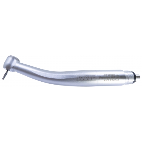
Premium LED 405 nm UV F.A.C.E. Handpiece — Fluorescence‑Aided Caries Excavation.
For dental clinicians and procurement specialists, precision and speed matter. JinDELL's F.A.C.E. handpiece uses 405 nm UV fluorescence to make carious dentin glow orange-red, while healthy enamel appears green—enabling minimally invasive excavation and safeguarding tooth structure. Supported by a built-in LED generator, 4-nozzle water cooling, and included safety goggles, this handpiece brings cutting-edge caries detection into routine practice.
-
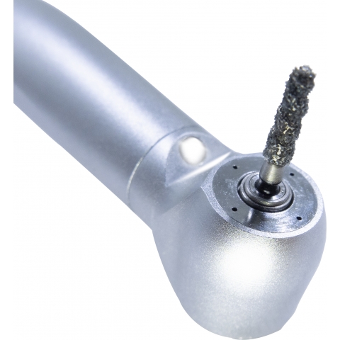
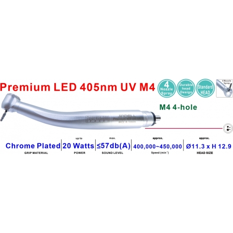
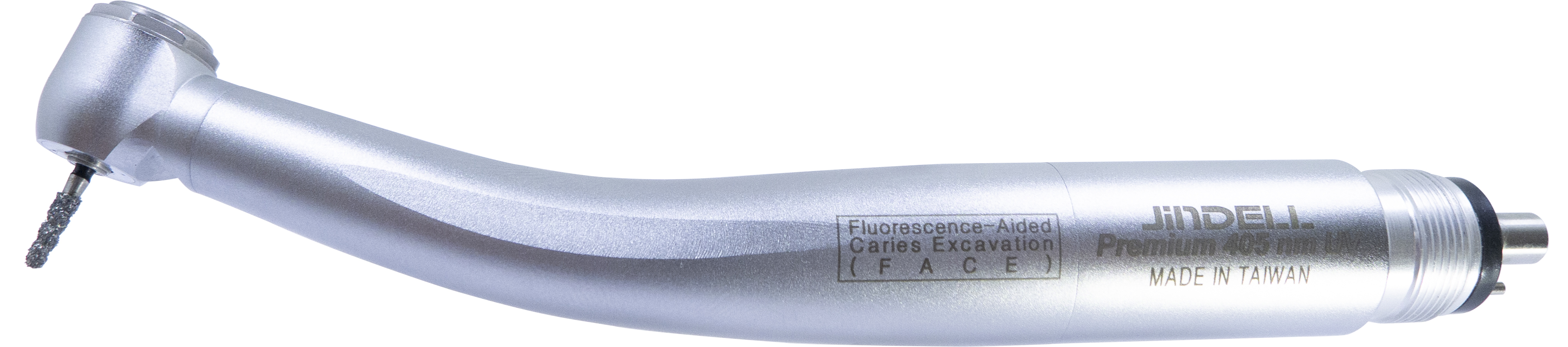
Built-in 405 nm UV LED generator integrated into the handpiece—no external cables needed.
-
4-nozzle water spray for efficient cooling during use.
-
The anti-retraction valve maintains hygiene by protecting the handpiece internals.
-
Includes protective UV goggles for clinician safety during use.
-
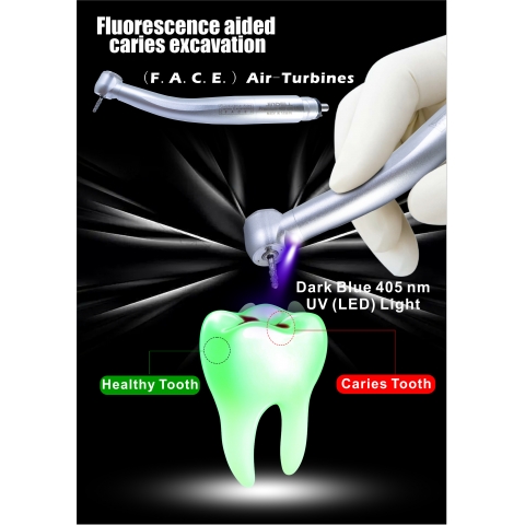
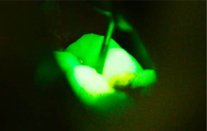
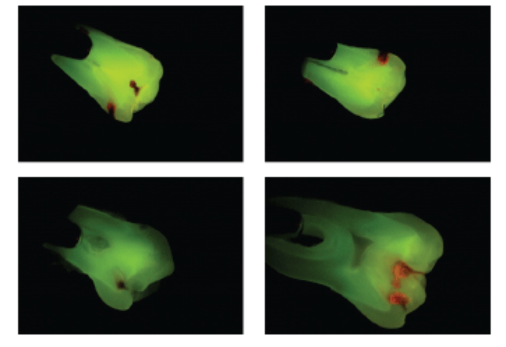
Fluorescence-led excavation: carious dentin (orange-red) vs healthy tissue (green), enabling minimally invasive removal and superior restorative visual margins.
-
Faster, more reliable caries removal: proven to reduce excavation time and improve accuracy over conventional methods.
 Built with an LED generator high-speed pneumatic handpiece featuring a four-nozzle water spray, it will also include a Dark Blue UV light for fluorescence checking.
 It will be shipped with goggles to check the fluorescence.
tay khoan nha khoa, 歯科用ハンドピース, 치과용 핸드피스, သွားနှင့်ခံတွင်းလက်ကိုင်, ด้ามจับทันตกรรม, Alat Tangan Pergigian, Handpiece ແຂ້ວ, Handpiece Gigi,ডেন্টাল হ্যান্ডপিস
Technical Specifications Comparison Table
Specification F.A.C.E. Handpiece UV Light Wavelength 405 nm (UV) LED Cooling 4-nozzle water spray Hygiene Anti-retraction valve included Safety Includes UV protective goggles Illumination Purpose Fluorescence caries vs healthy tissue Clinical Benefit Minimally invasive removal, precision Innovation Integrated UV generator with no external bulbs
How It Works & Clinical Benefits
-
Mechanism: Bacterial byproducts (porphyrins) in carious dentin fluoresce orange-red under 405 nm UV; healthy tooth structure glows green.
-
Benefit #1: Only decayed tissue is removed, preserving healthy enamel/dentin and reducing cavity size and tooth strength loss.
-
Benefit #2: Restoration margins are clearly visible without dyes or imaging—saving chair time and enhancing accuracy.
-
Scientific Support: Studies show F.A.C.E. improves the accuracy of decay removal and reduces the risk of residual caries, all while not significantly enlarging cavity size.
Hygiene, Sterilization & Safety
-
Non-contaminating design: anti-retraction valve protects against backflow.
-
Sterilization: Follow regular handpiece sterilization procedures—avoid direct exposure of UV components; however, the outer shell is compatible with standard procedures.
-
Safety Gear: UV-protective goggles are included to shield clinicians during fluorescence use.
CTAs (Effective Calls to Action)
-
Request a F.A.C.E. Handpiece Demo
-
Get Bulk Pricing for Wholesalers
-
Download Scientific Brochure & Clinical Case Studies
-
Become a Distributor or Supplier
-
Contact JinDELL for Clinical Training / Support
FAQ (English)
Q1. What does “F" A.C.E.” "mean?
It stands for Fluorescence-Aided Caries Excavation, utilizing 405 nm UV light to distinguish between caries and healthy tooth structure.
Q2. How is carious dentin distinguished?
Healthy enamel glows light green under the UV light, while its orange-red fluorescence is visible.
Q3. Are safety measures included?
Yes—the handpiece ships with UV-safe protective goggles for clinical safety.
Q4. Does it shorten procedure time?
Yes. It simplifies caries detection and excavation by eliminating dye and visual imaging steps, thereby improving efficiency.
Q5. Does it support minimally invasive dentistry?
Exactly—only infected dentin is removed, preserving healthy tooth tissue.
Q6. How does it impact restorative work?
UV light makes restorative materials clearly visible against enamel, aiding margin detection and reducing over-preparation.
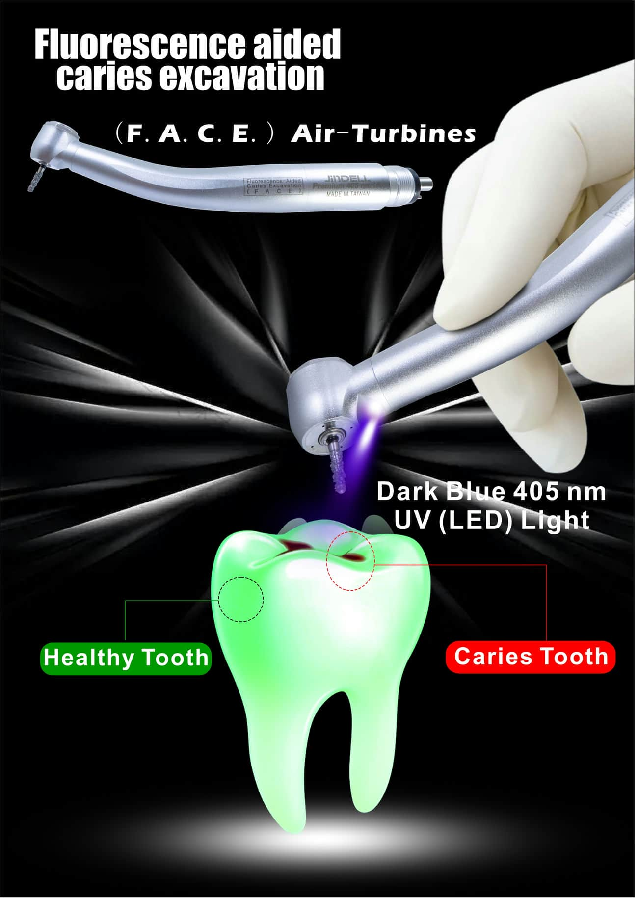
- Dental professionals using fluorescence-aided caries excavation (F.A.C.E.) turbines will find that removing infected dentin is easier and quicker than with conventional methods.
- These Air Turbines emit high-intensity 405 nm (UV) light. LED light is an alternative to white light, which can dramatically enhance the visualization of caries and restorative materials.
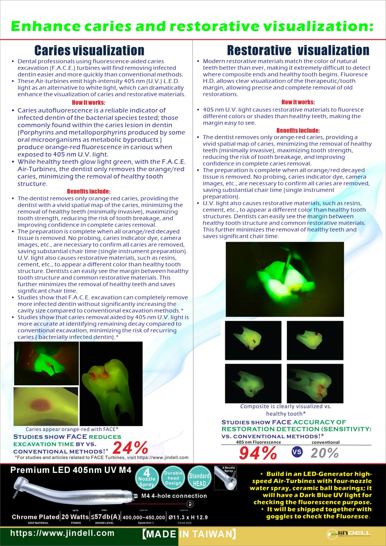
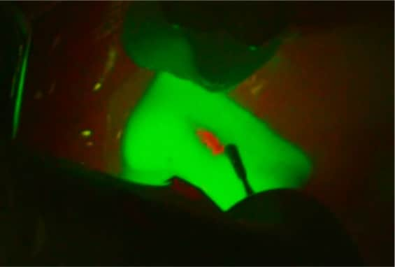
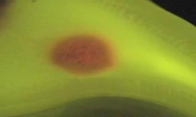
- Caries autofluorescence is a reliable indicator of infected dentin, as it detects the bacterial species tested; those commonly found within the caries lesion in dentin (Porphyrins and metalloporphyrins produced by some oral microorganisms as metabolic byproducts) produce orange-red fluorescence in carious dentin when exposed to 405 nm UV light.
- While healthy teeth glow light green, with the F.A.C.E. Air-Turbines, the dentist only removes the orange/red caries, minimizing the removal of healthy tooth structure.
- The dentist removes only orange-red caries, providing the dentist with a vivid spatial map of the caries, minimizing the removal of healthy teeth (minimally invasive), maximizing tooth strength, reducing the risk of tooth breakage, and improving confidence in complete caries removal.
- The preparation is complete when all orange/red decayed tissue is removed. No probing, caries indicator dye, camera images, etc., are necessary to confirm that all caries have been removed, saving substantial chair time (single-instrument preparation).
- UV light also causes restorative materials, such as resins and cements, to appear a different color than healthy tooth structure. Dentists can easily see the margin between healthy tooth structure and common restorative materials. This further minimizes the removal of healthy teeth and saves a significant amount of chair time.
- Studies show that F.A.C.E. excavation can completely remove more infected dentin without significantly increasing the cavity size compared to conventional excavation methods.*
- Studies show that caries removal aided by 405 nm UV light is more accurate at identifying remaining decay compared to conventional excavation, thereby minimizing the risk of recurring caries (bacterially infected dentin).*
*Studies show F.A.C.E. reduces excavation time by vs. conventional methods: 24%
- Modern restorative materials closely match the color of natural teeth, making it extremely difficult to detect where the composite ends and the healthy tooth begins. Fluoresce H.D. allows clear visualization of the therapeutic/tooth margin, allowing precise and complete removal of old restorations.
- 405 nm UV light causes restorative materials to fluoresce different colors or shades than healthy teeth, making the margin easy to see.
- The dentist removes only orange-red caries, providing a vivid spatial map of the caries, thereby minimizing the removal of healthy teeth (minimally invasive) and maximizing tooth strength, which reduces the risk of tooth breakage and improves confidence in complete caries removal.
- The preparation is complete when all orange/red decayed tissue is removed. No probing, caries indicator dye, camera images, etc., are necessary to confirm that all caries have been removed, saving substantial chair time (single-instrument preparation).
- UV light also causes restorative materials, such as resins and cements, to appear a different color than healthy tooth structures. Dentists can easily see the margin between healthy tooth structure and common restorative materials. This further minimizes the removal of healthy teeth and saves a significant amount of chair time.
Studies show FACE ACCURACY OF RESTORATION DETECTION (SENSITIVITY) vs. conventional methods:*

University-Based Scientific Evidence